Laboratory of Photosynthesis
Radek Kana`s group
Membranes heterogeneity in photosynthesis
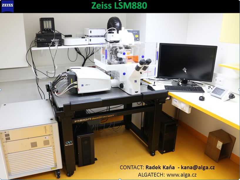
Confocal Microscopy
 |
BASIC DESCRIPTION:
Our laboratory operates high-sensitivity/high-speed confocal Laser Scanning Microscope (LSM Zeiss 880) with standard features (for cell biology); the instrument is additionally optimized for photosynthetic microrganisms and chloroplasts with their high autofluorescence. The microscope is suitable for standard 2D, 3D imaging and time lapse image acquisition, advanced microscopic methods (FRAP, FCS). and super-resolution (AiryScan). The instrument contained all necessary objectives (oil objectives NA = 1.4 and NA=1.45 with 63x and 100x magnification respectively; water NA=1.4 - 40x magnification objective for FCS). There are 7 laser lines (405, 458, 488, 543, 594 and 633 nm) and variable detection by 2 PMT detectors and spectral detector (cells spectrum in one image, 8nm step, 32 channels, speed 5 fps), transmission and super-resolution detector (Airyscan) with high signal to noise ration and 120 nm spatial resolution in x-y [1].
[1] L. Scipioni, L. Lanzanó, A. Diaspro and E. Gratton, Nature Communications, 2018, 9, 5120.
APPLICATIONS:
The laboratory confocal is suitable for superresolution imaging of all classical fluorescence dyes and proteins excitable by our laser set (e.g. DAPI, TFP, GFP, Alexa, CFP and others) used in single-cell biology. Additionally it allows measurement of autofluorescence of chlorophylls and various phycobiliproteins from different organisms (algae, cyanobacteria). Experimental routines include methods for detectionof proteins mobility and interaction at the nanoscale level including photoactivation methods (FRAP – Fluorescence Recovery After Photobleahcing), correlation methods (e.g. FCS – Fluorescence Correlation Spectroscopy). The software allows 3D reconstirution of Z-stack imaging in standard (300 nm in x-y) and in superesolution mode (120 nm in x-y).
|
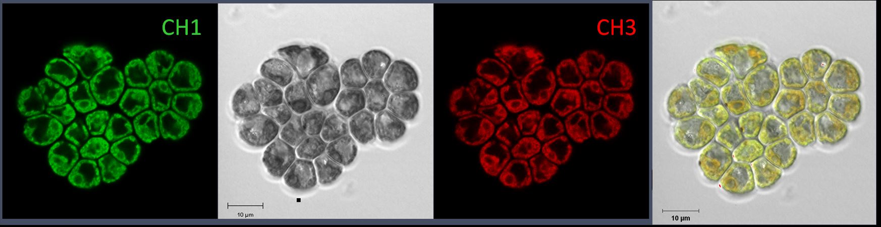
Transmission and autofluorescence image of green algae Chloromonas Brevispina |
|
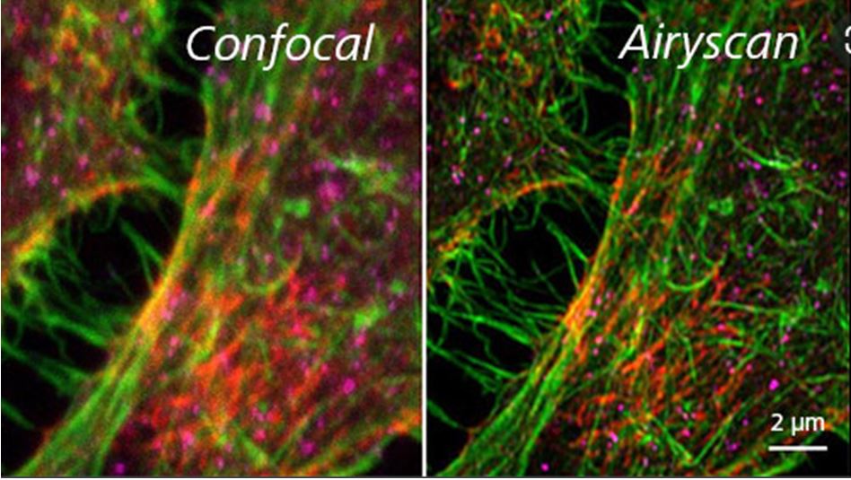
Cells stained for actin (green), septin A (red) and adapter protein AP-3 (magenta) Image courtesy of S. Traikov, BIOTEC, TU Dresden, Germany |
|
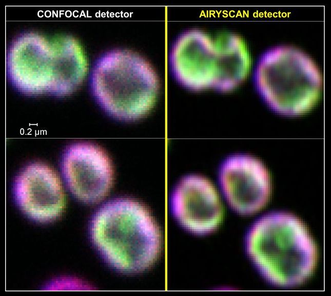
Superresolution of cyanobacteria cells thylakoids (GREEN – YFP tagged Photosystem I) |
|
3D reconstitution of Chromera velia chloroplast |
|
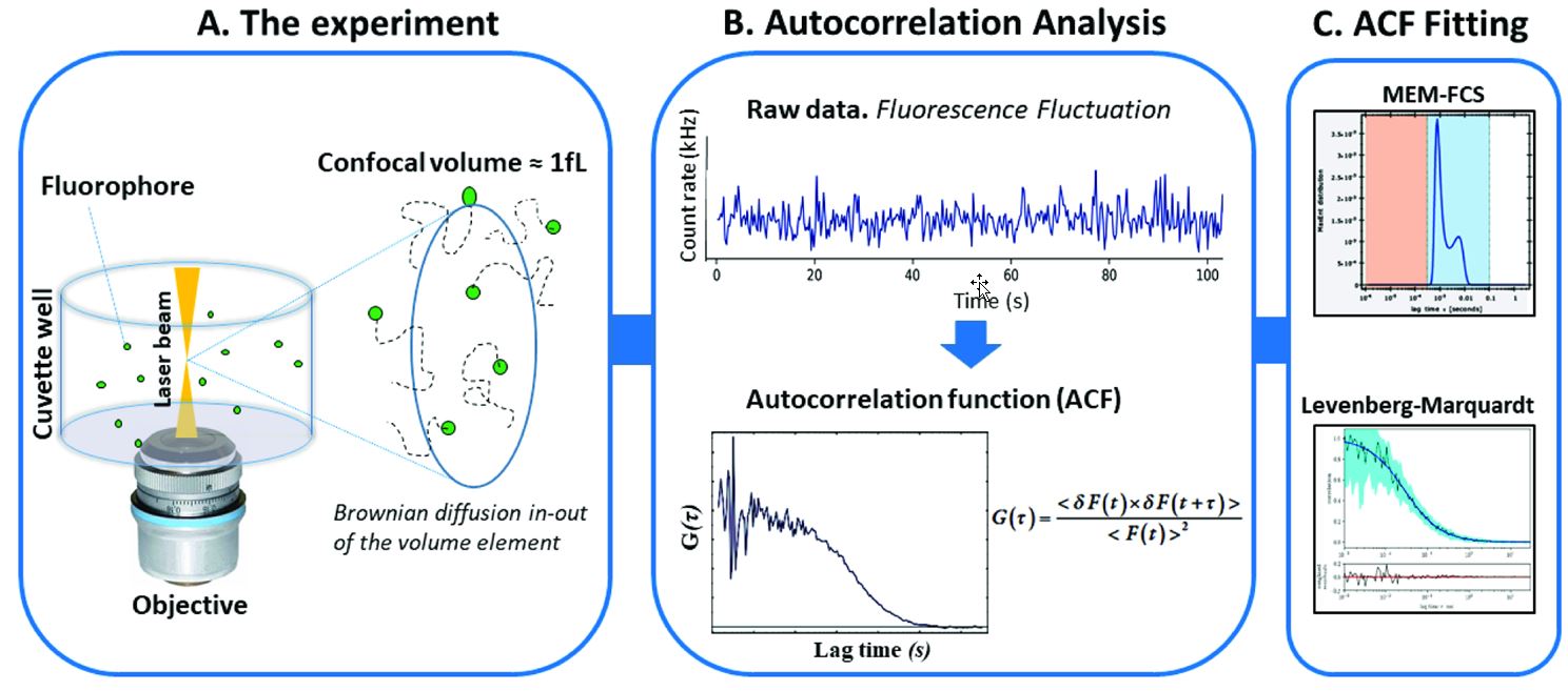
The Figure illustrates the workflow in a typical FCS experiment for characterization of protein sizes, oligomerization and diffusion |
|
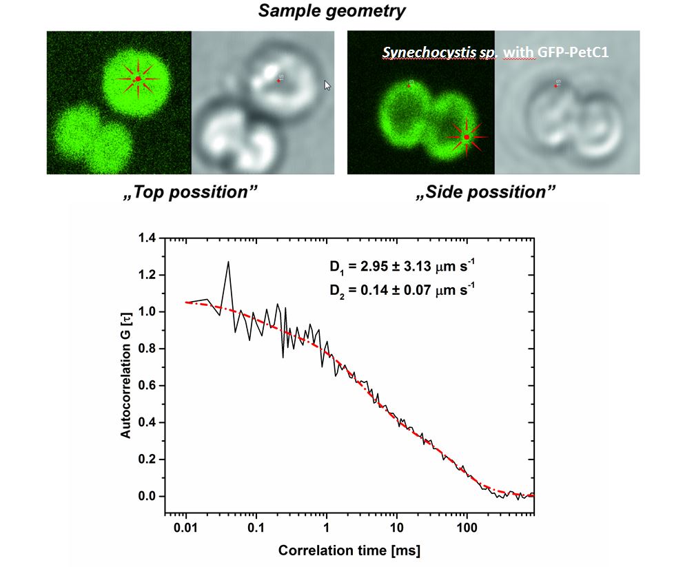
Example of FCS application for detection of PetC1-GFP protein diffusion in thylakoids (see Kaňa et al. Life 2021, 11) |
Contact:
Radek Kaňa kana@alga.cz
Barbora Šedivá sediva@alga.cz