Laboratory of Photosynthesis
Josef Komenda`s group
Biogenesis of Photosystem II
Research Projects
The Role of FtsH Proteases in Biogenesis of Thylakoid Membranes and Photosynthetic Complexes in Cyanobacteria
Funded by the Grant Agency of the Czech Republic - Junior Project, 2020-2022.
Principal investigator: Vendula Krynická, Ph.D.
FtsHs are universally conserved transmembrane metalloproteases. In plants, they are key players in ongoing photosynthesis and an early stage of plastid development. In cyanobacteria, FtsHs are mostly known for PSII repair during photoinhibition. Our preliminary results, however, indicate that cyanobacterial FtsHs are also involved in assembly of photosynthetic complexes. They were repeatedly copurified with PSII and PSI reaction centres as well as proteins involved in chlorophyll biosynthesis and thylakoid membrane (TM) formation. Moreover, suppression of the essential FtsH1/3 complex causes aberrant TM architecture.
Like other cyanobacteria, the model organism Synechocystis PCC6803 contains four FtsH homologues, FtsH1-FtsH4. In vivo, they form two hetero-oligomeric complexes, FtsH1/3 and FtsH2/3, and one homo-oligomeric complex, FtsH4. Our previous studies brought new information about the architecture, composition and localization of the FtsH complexes in vivo. Recent results from confocal microscopy of GFP tagged FtsH derivatives indicate that the most abundant FtsH2/3 complex is located in the thylakoid membrane, whereas the less abundant essential FtsH1/3 is located in plasma membrane. The FtsH4 homo-oligomeric complex was previously localized within the TM. (Komenda et al., 2006; Boehm et al., 2012; Krynická et al., 2014; Krynická et al., 2015)
The aim of the project is to specify the role of FtsHs in PSII and PSI de novo assembly and TM biogenesis. We focus on FtsHs localization and the effect of FtsH deficiency and overexpression on the photosystem assembly under conditions inducing TM biogenesis (regreening). We also isolate sections of the TM enriched by FtsH complexes and characterize their protein composition. Additionally, we would like to further specify the structure of purified FtsH complexes.
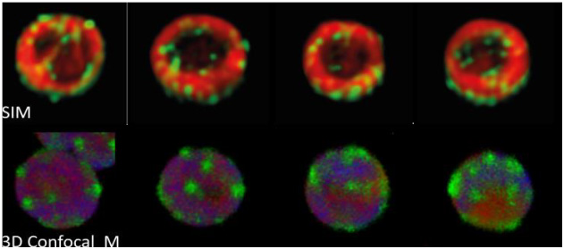
Localization of FtsH4 using fluorescence signal of FtsH4-GFP, chlorophyll and phycobilins. Upper: 3D Structured illumination microscopy (SIM). 3D image of cell cross-sections made of 2D axial slices 125 nm apart. Lower: 3D confocal microscopy image is made of 15 2D slices with a thickness of 150 nm. red - Chlorophyll (Chl); green - GFP; yellow - Overlap of Chl with GFP; blue - Phycobilins; violet - Overlap of Chl with Phycobilins.
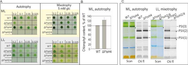
Phenotype of Synechocystis strains missing thylakoid FtsH. A: Growth assay of WT, ΔFtsH2, ΔFtsH4 and ΔFtsH2/4 mutants on agar plates exposed to moderate light (ML, 50 μE)/ low light (LL, 5 μE) and autotrophic or mixotrophic conditions. B: Content of chlorophyll in ΔFtsH4 as a percentage of WT levels of Chl, mean of six measurements. C: Separation of PS complexes in WT, ΔFtsH2, ΔFtsH4 and ΔFtsH2/4 mutants using CN PAGE. Loaded samples were normalized per number of cells; PSI(3): PSI trimer, PSII(1) and or PSII(2): PSII monomer or dimer, respectively. Chl fl: chlorophyll auto-fluorescence at room temperature.